Plant Cell Cytokinesis Microscope : Biology Free Full Text Mitotic Spindle Assembly In Land Plants Molecules And Mechanisms Html / While this is the last event of cell division, it starts early on during the anaphase stage of mitosis in most eukaryotic cells.
Plant Cell Cytokinesis Microscope : Biology Free Full Text Mitotic Spindle Assembly In Land Plants Molecules And Mechanisms Html / While this is the last event of cell division, it starts early on during the anaphase stage of mitosis in most eukaryotic cells.. 4 1 understanding mitosis lessons tes teach. What feature will indicate that it has just reached the end of interphase? Plant mitosis wall chart with plastic edging for hanging amazon. Most see will be in interphase (between cell divisions). • quadruple stain allium microscope slides provide excellent contrast between the cell wall, nuclear membrane, and the chromosomes.
But at the same time it is interpretive. Many items at sale prices. B0007563 mitosis in onion root cells biologia celular ciclo. Here's a diagram of a plant cell: In part i, you observed cell division in animal cells.
Cytokinesis Png Images Pngwing from w7.pngwing.com Cell wall, nuclei, chromatin, cytoplasm and cell plate. Draw a cell that represents cytokinesis in onion root tip and label with the following terms: During cytokinesis, a cell separates the two nuclei, formed during the later stages of cell division i.e. So, this is the key difference between plant and animal cytokinesis. Focus at 100x and re center so that you are focused on the more 'square' meristem cells. B0007563 mitosis in onion root cells biologia celular ciclo. Observing mitosis in plant cells continued 3 21 linn ientii n ll igts eserve • a 100x oil immersion lens is a useful tool to visualize spindle fibers and cell plates. General mechanisms of cytokinesis in eukaryotes while the process.
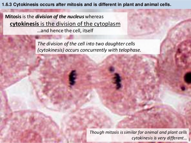
Cytokinesis is the second part of the mitotic phase during which cell division is completed by the physical separation of the cytoplasmic components into two daughter cells.
The cell more information cell wall atlas of plant and animal. If a cell undergoes mitosis but does not complete cytokinesis, it would become a cell with _____ nuclei. Metaphase of an onion root tip cell under the microscope stock. Plant mitosis wall chart with plastic edging for hanging amazon. Imagenes fotos de stock y vectores sobre mitosis of plant. The diagram is very clear, and labeled; What feature will indicate that it has just reached the end of interphase? This cell is most likely a plant cell in the process of cytokinesis. During the cytokinesis phase of cell division, plant cell division, plant cells develop a cell plate instead of a cleavage furrow. Cytokinesis refers to the division of the cytoplasm during cell division (mitosis). Focus at 100x and re center so that you are focused on the more 'square' meristem cells. • quadruple stain allium microscope slides provide excellent contrast between the cell wall, nuclear membrane, and the chromosomes. So, this is the key difference between plant and animal cytokinesis.
General mechanisms of cytokinesis in eukaryotes while the process. The stages of mitosis and cell division. There are, however, differences in the process in plant cells and in animal cells, which can be seen when the phases of mitosis and cytokinesis are studied. In plant cells cytokinesis is accomplished by the formation of a. Cytokinesis is the second part of the mitotic phase during which cell division is completed by the physical separation of the cytoplasmic components into two daughter cells.
Ib Biology 1 6 1 1 Slides Mitosis Stem Cells from image.slidesharecdn.com The stages of mitosis and cell division. Cytokinesis • after the plant cell separates the sister chromatids and builds new nuclear membranes to create two nuclei, it divides its cytoplasm into two parts by forming new plasma membrane and cell wall down the middle of the cell. During cytokinesis in animal cells, a ring of actin filaments forms at the metaphase plate. Produkte für gewerbe und wissenschaft. In contrast, animal cell cytokinesis occurs via the formation of a cleavage furrow. Top the purpose of this page is to show interested individuals how mitosis occurs in a stamen hair cell of the spiderwort plant, tradescantia virginiana. Metaphase of an onion root tip cell under the microscope stock. (a) a drawing of cell walls from the cork tissue of an oak (quercus sp.) tree, published.
• quadruple stain allium microscope slides provide excellent contrast between the cell wall, nuclear membrane, and the chromosomes.
So, this is the key difference between plant and animal cytokinesis. This cell is most likely a plant cell in the process of cytokinesis. Using your microscope, scan the slides to In animal cells, a ring of actin fibers is formed around the periphery of the cell at the former metaphase plate (cleavage furrow). In plant cells, golgi vesicles coalesce at the former metaphase plate, forming a phragmoplast. During the cytokinesis phase of cell division, plant cell division, plant cells develop a cell plate instead of a cleavage furrow. Through a microscope, you can see a cell plate beginning to develop across the middle of a cell and nuclei forming on either side of the cell plate. • quadruple stain allium microscope slides provide excellent contrast between the cell wall, nuclear membrane, and the chromosomes. Although the stages of mitosis are similar for most eukaryotes, the process of cytokinesis is quite different for eukaryotes that have cell walls, such as plant cells. Set up your microscope, place the onion root slide on the stage and focus on low (40x) power. Imagenes fotos de stock y vectores sobre mitosis of plant. Plant growth and development relies on the accurate positioning of the cell plate between dividing cells during cytokinesis. Metaphase of an onion root tip cell under the microscope stock.
In higher plant cells, cytokinesis is regulated by the cell wall and occurs by a different mechanism. Imagenes fotos de stock y vectores sobre mitosis of plant. In thissection, you will observe the process in plant cells. Plant mitosis wall chart with plastic edging for hanging amazon. Cytokinesis is the second part of the mitotic phase during which cell division is completed by the physical separation of the cytoplasmic components into two daughter cells.
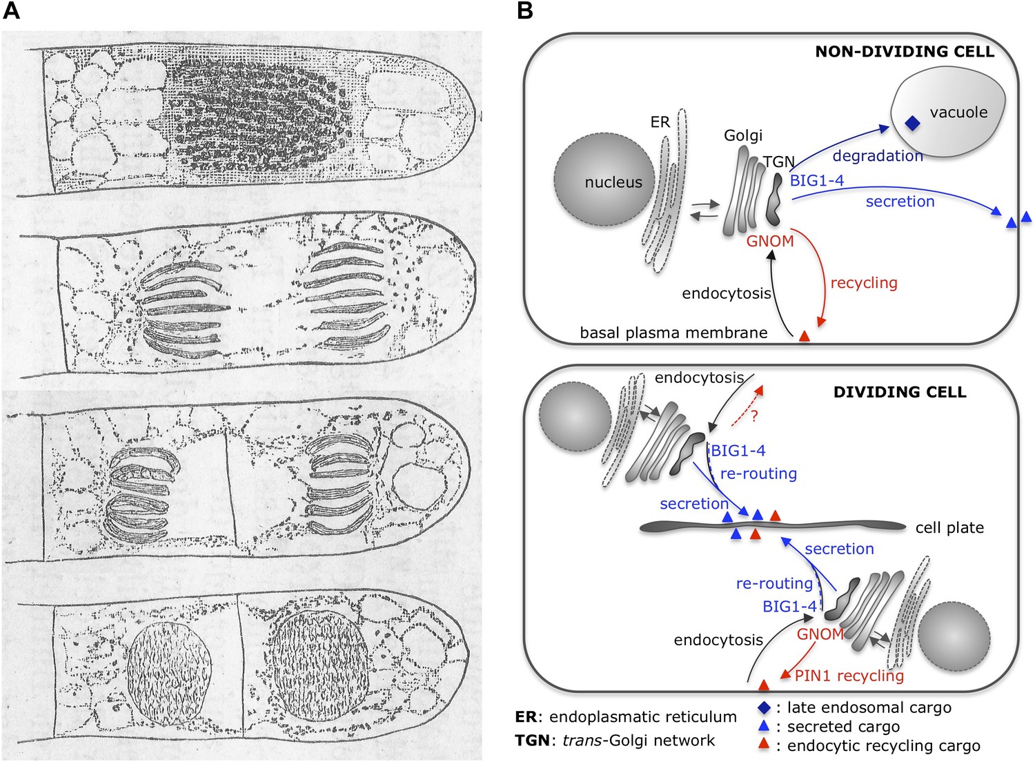
Plant Cytokinesis Illuminating Traffic Control For Cell Division Planes Elife from iiif.elifesciences.org While this is the last event of cell division, it starts early on during the anaphase stage of mitosis in most eukaryotic cells. Plant growth and development relies on the accurate positioning of the cell plate between dividing cells during cytokinesis. By cytokinesis summary plants, people and the environment: When observed in the optical microscope, the midbody reveals a dense matrix of tightly packed polar microtubules remaining from the mitotic spindle. We combined high spatial and tempor … In thissection, you will observe the process in plant cells. Move your slide so that your field of view is centered on the root tip. This cell is most likely a plant cell in the process of cytokinesis.
Through a microscope, you can see a cell plate beginning to develop across the middle of a cell and nuclei forming on either side of the cell plate.
(a) a drawing of cell walls from the cork tissue of an oak (quercus sp.) tree, published. Sketch and label cytokinesis of plant cell brainly in. There are very few similarities between animal cell and plant cell cytokinesis. General mechanisms of cytokinesis in eukaryotes while the process. Many items at sale prices. Even though the cells in these tissues are rapidly dividing, of the cells you. Plant mitosis wall chart with plastic edging for hanging amazon. In animal cells, a ring of actin fibers is formed around the periphery of the cell at the former metaphase plate (cleavage furrow). In plant cells cytokinesis is accomplished by the formation of a. Unit 1 cell molecular biology cell growth cell cycle ppt. When observed in the optical microscope, the midbody reveals a dense matrix of tightly packed polar microtubules remaining from the mitotic spindle. A cell is observed under the microscope. The plant cell and the cell cycle cells and microscopy.
During the cytokinesis phase of cell division, plant cell division, plant cells develop a cell plate instead of a cleavage furrow plant cell cytokinesis. • the cytoskeleton moves small vesicles containing cell wall material into a line in the middle of the cell.


0 Comments